Category Archives: Archaeology

Building the Crypt

A large quantity of human remains were uncovered during the archaeological excavation in 2006. Since then they have been the subject of a great deal of study and research, some of which is ongoing. However, the aim has always been to re-inter them within the building. This is easy to write but more difficult to do, not least because of the large volume the bones occupy. It is also important that wherever they are buried it is in a place where they will not be disturbed again. Our architect was asked to draw up plans for a crypt to be built below the floor level of the lowest level of the proposed 4-storey building within the shell of the former East Kirk, in other words below the lowest levels of the dig.

We were able to obtain funding from the Town Centre Fund, a Scottish Government initiative being administered locally by Aberdeen City Council and we are very grateful to them for this. Once all permissions were obtained the contract to build the crypt was signed with CHAP Construction of Westhill. Work was scheduled to start in March 2020 – just after the beginning of the first Covid-19 lockdown. Not surprisingly it had to be postponed, finally getting underway in September 2020.

Early stages of excavation
Getting deeper

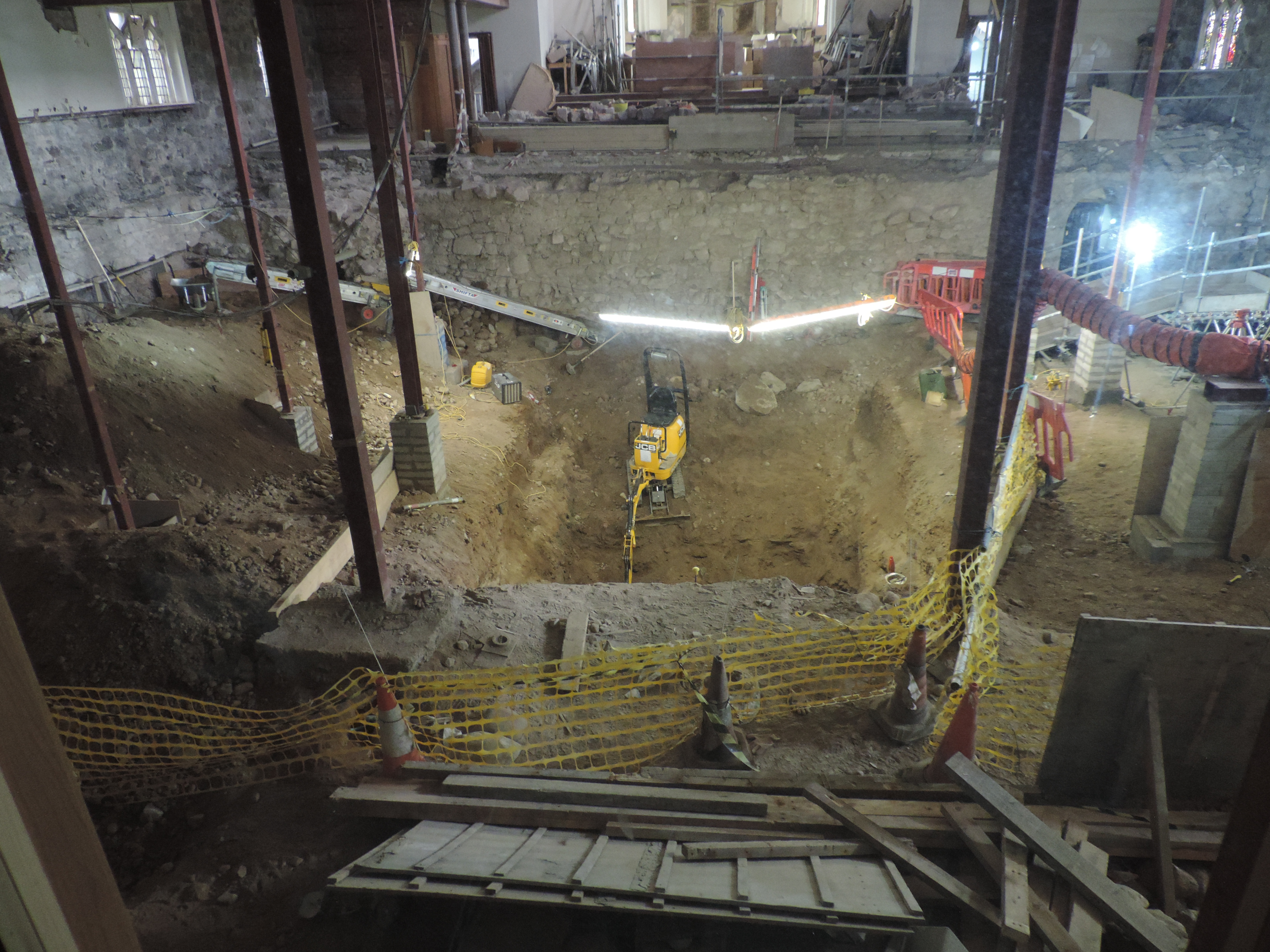
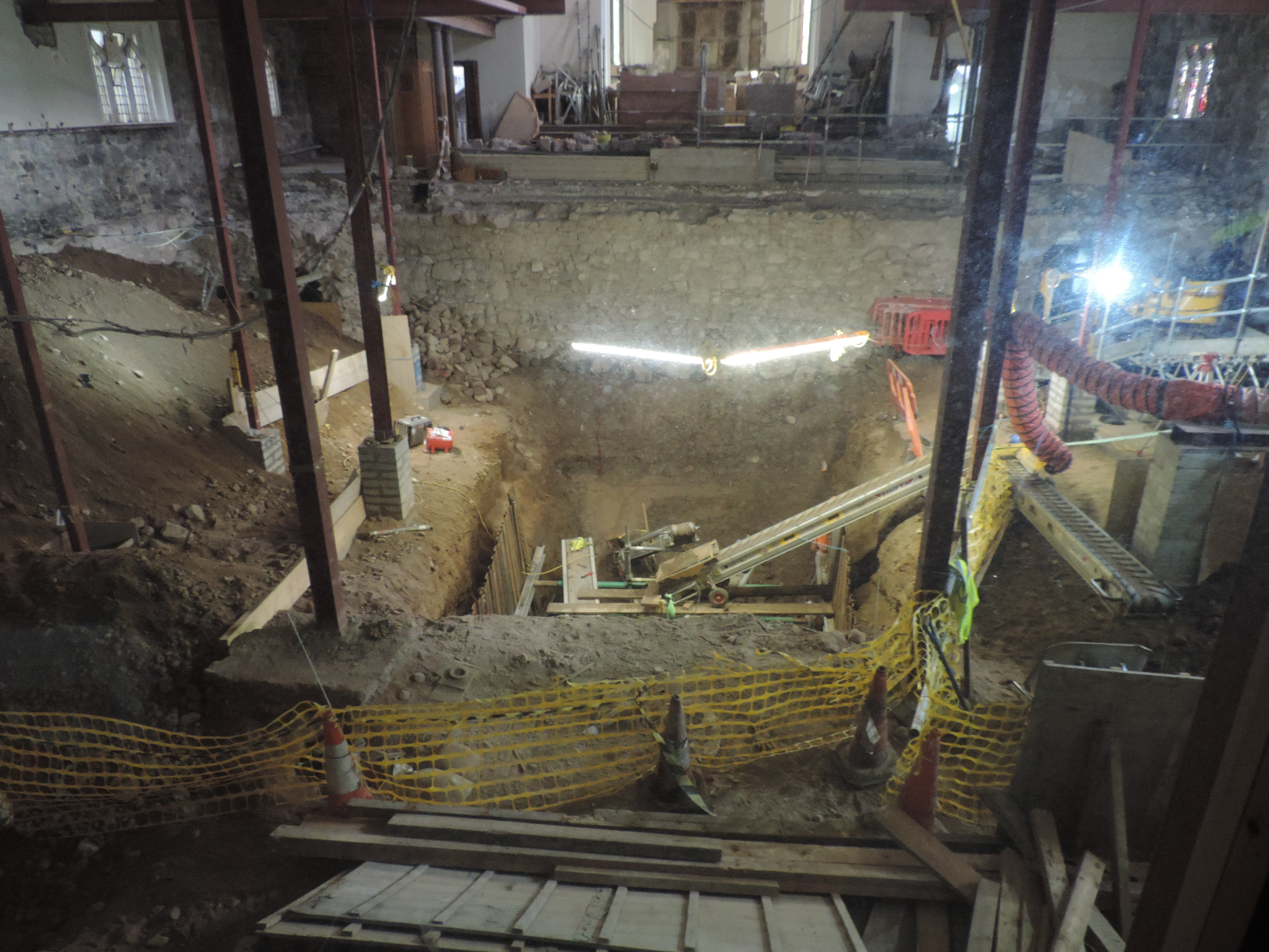
The first and slowest part of the work was the excavation of the large hole required. This was about 25 feet long and 11 feet wide and went down about 7 feet. Special machinery was needed which required a new temporary ramp to be built to give access.

The base is put in place
The walls are built

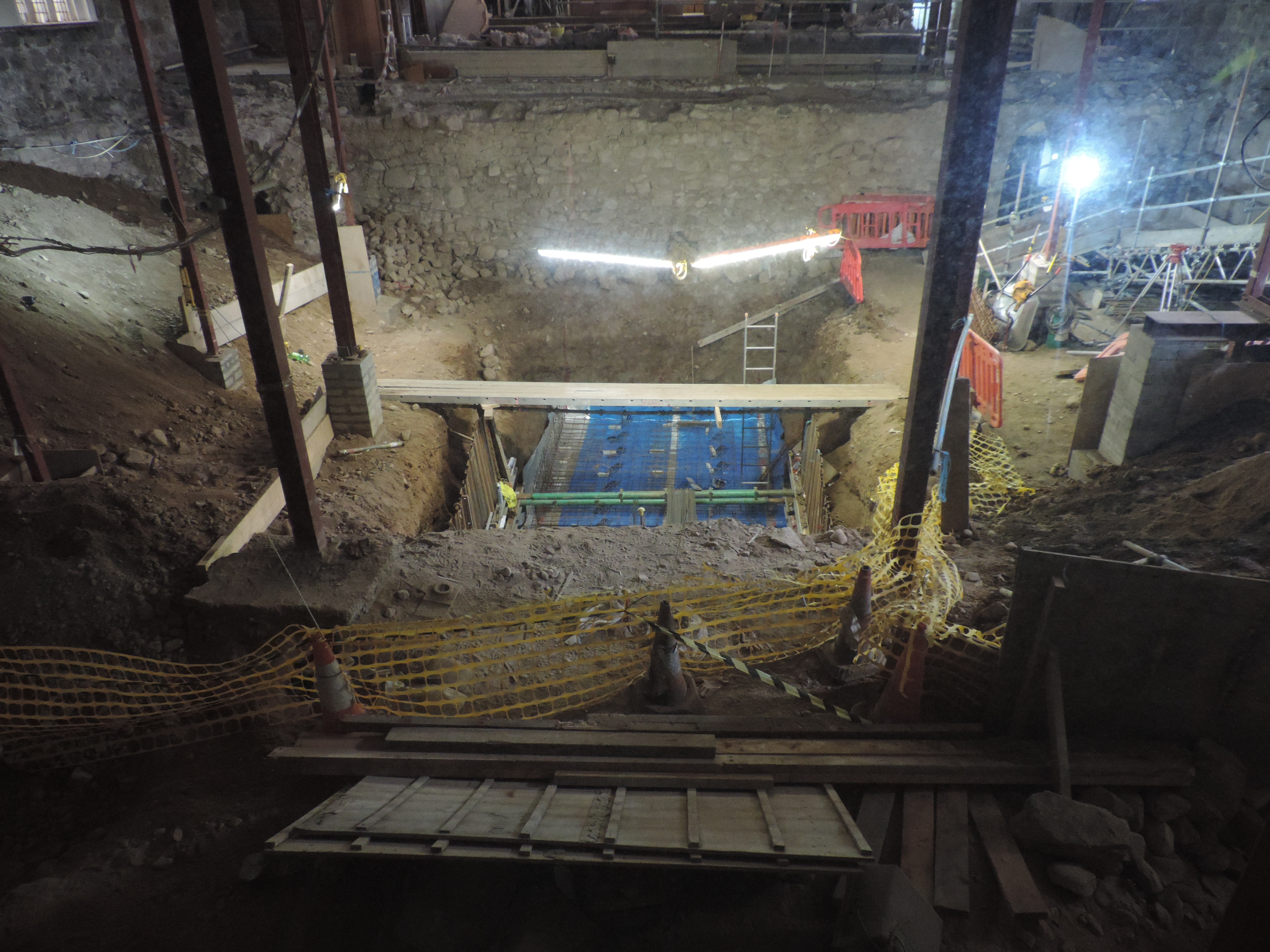
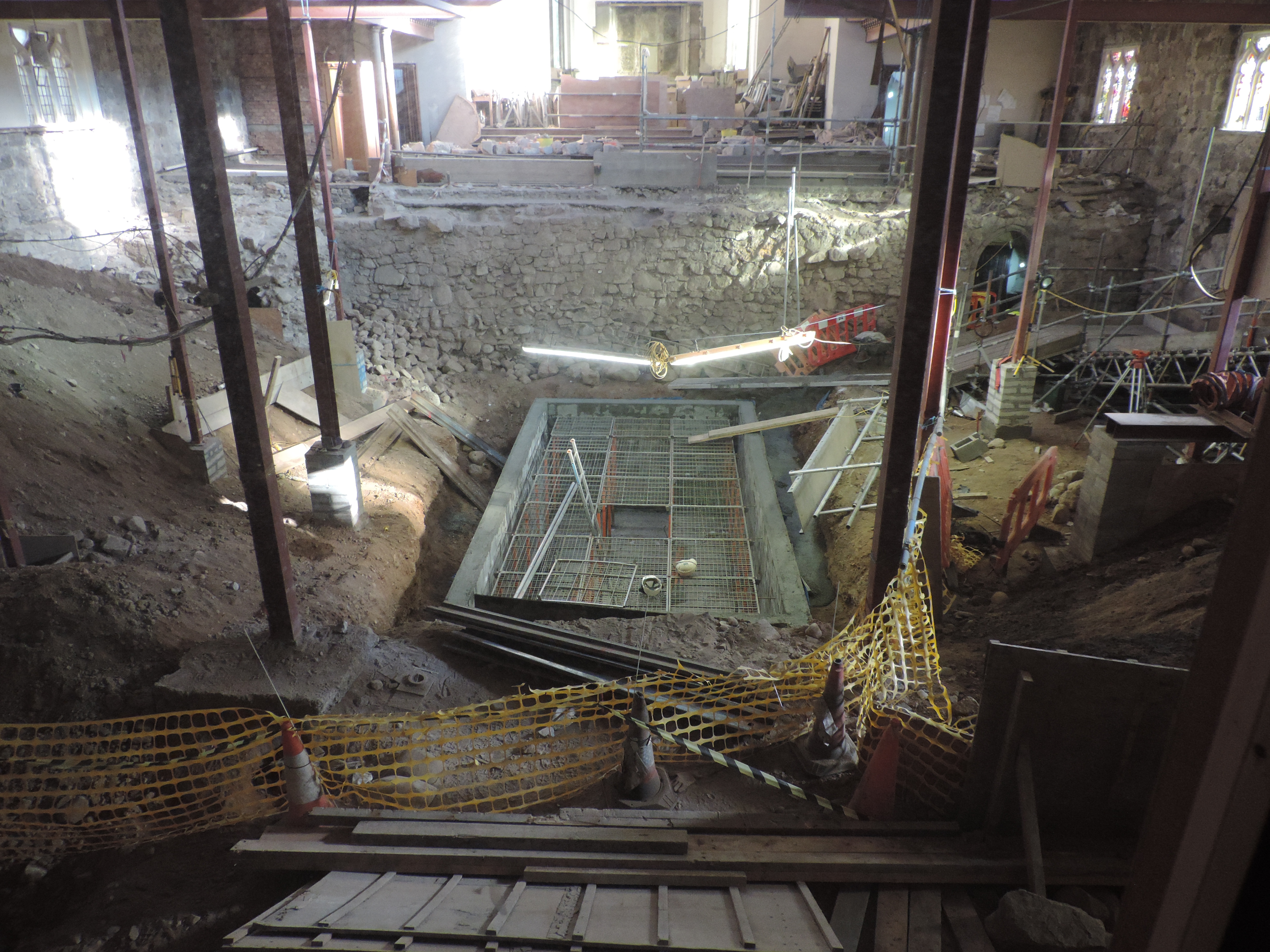
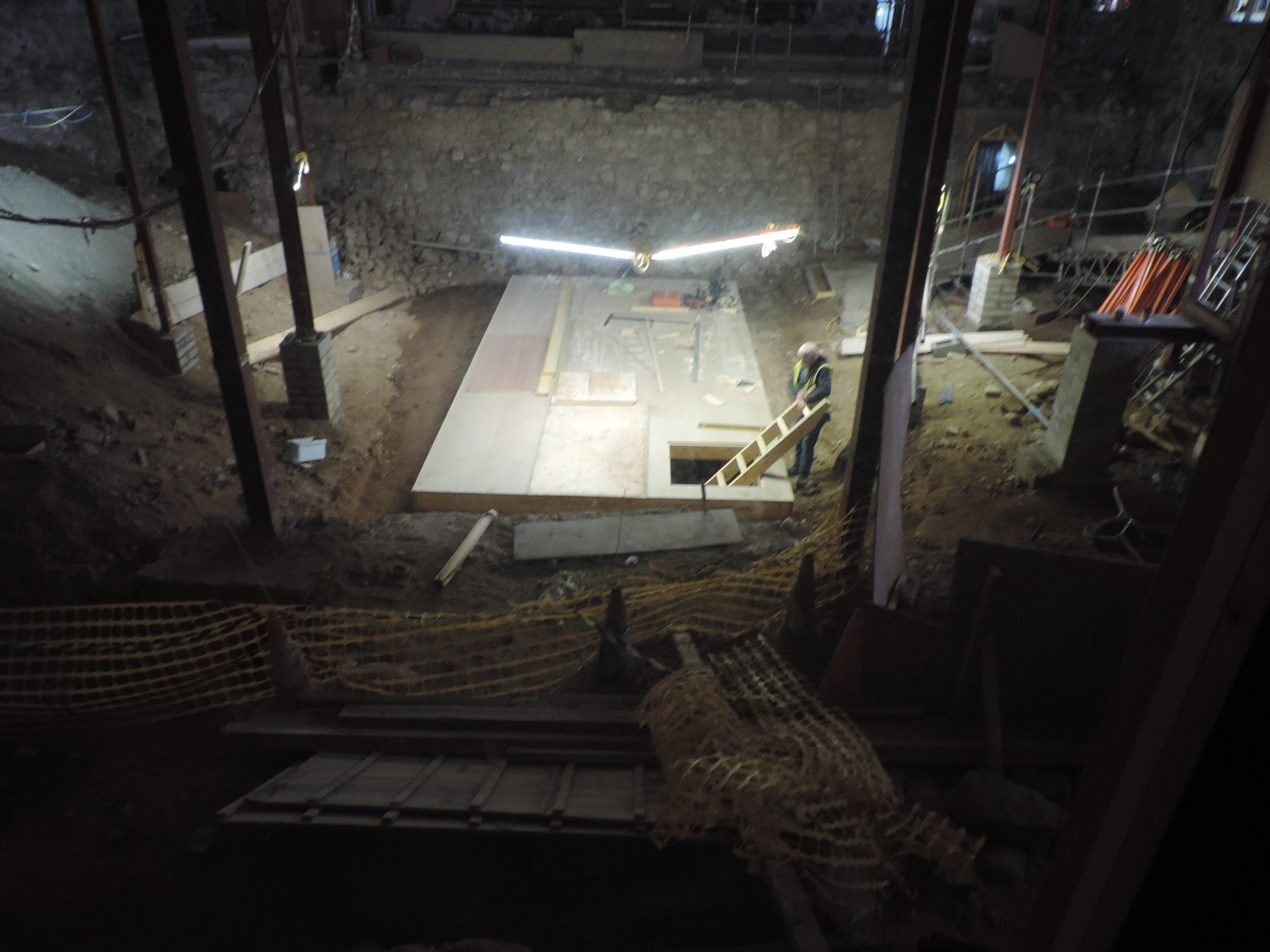
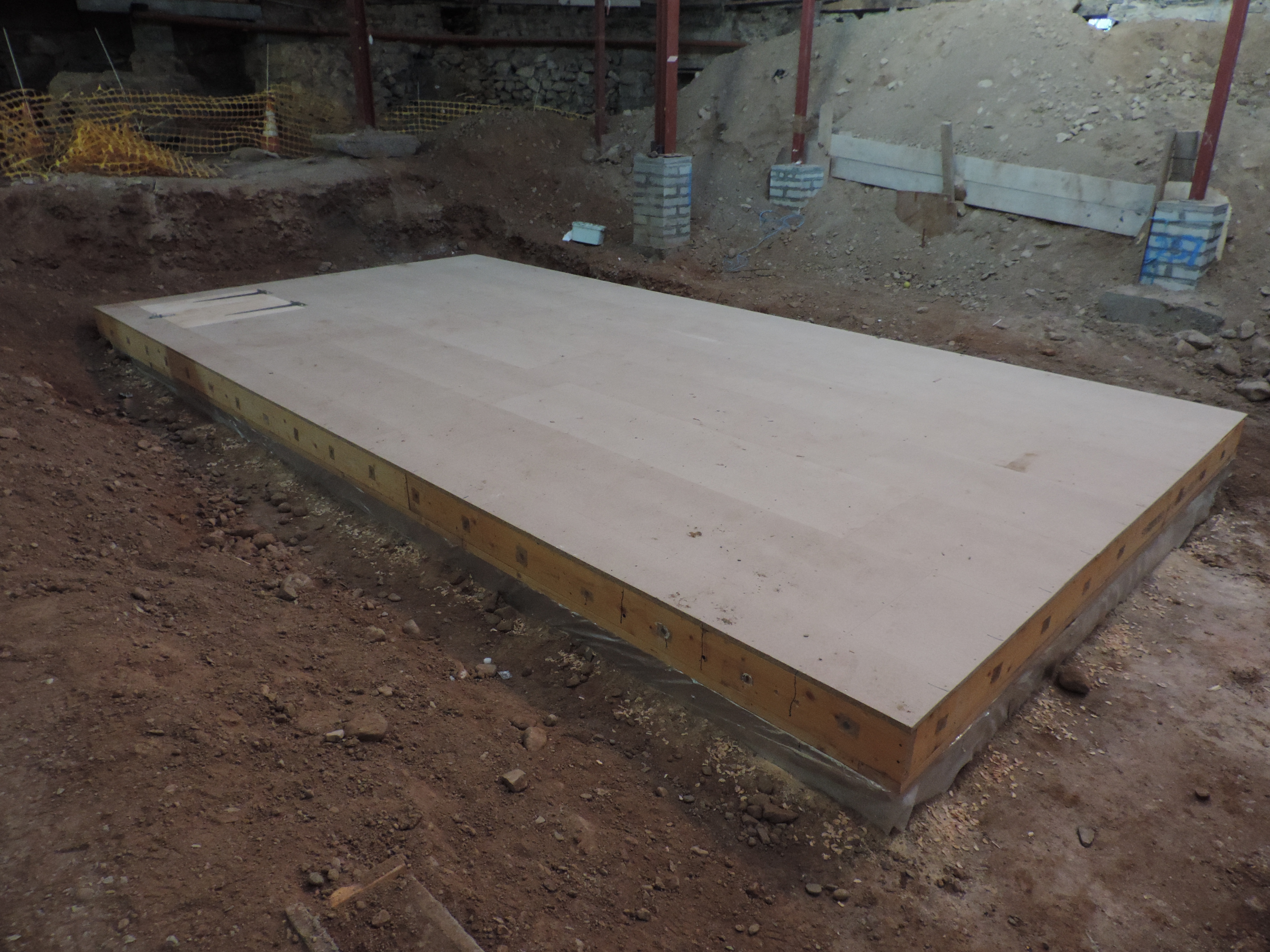
Once excavation was complete, the concrete base was made, then the walls were constructed and finally a wooden covering with access hatch and internal ladder was put in place. Work was completed in November. A number of photographs, mostly taken from the ‘viewing window’ off Drum’s Aisle, are included to show progress.

A wooden closure was made
Finished crypt – seen from the opposite end

At present none of the bones have been returned to the crypt because they all require placing into special boxes before this can be done. That will take quite a long time to process. Eventually, however, it is intended that there will be a religious ceremony to mark the laying to rest of the remains once more.

Worked stones

During the archaeological dig in 2006 a large number of worked stones were uncovered. On some stones, this working was quite small, for example one flat surface, or a rough piece with some chisel marks or a mason mark. Others had been carefully carved to produce a complex profile which had once been part of a building, but had since been discarded. One thing which became clear during the dig was that, as each new development of the building took place, the previous building was only partially demolished but not completely removed. It appears that some of the stone was either reused or it was left in situ.

A piece of shaped stone reused in a pillar base built in the late 1490s

The first photograph shows a piece of sandstone, probably from an earlier doorway, but reused in one of the 15th century pillar bases.

The next three photographs show a pillar column, first as it was being uncovered by the archaeologist, second once removed from the ground and third in comparison with one of the pillars in Drum’s Aisle

A slender broken pillar being excavated.
Part of the pillar once excavated


The excavated pillar in Drum’s Aisle beside a pillar


A carved stone, possibly from a doorway in an earlier building

The final photograph is of another carved piece of stone which may have been part of a door or archway in an earlier building. A detailed study of these stones has still to be completed but once done they could tell us a great deal about the appearance of the previous buildings.

(The photographs are copyright Aberdeen Art Gallery & Museums Collections and are used with permission)

Broken Bones

Archaeology is not just digging up bones and other artefacts. Sometimes, it can throw up some interesting ‘whodunit’ questions. This post reports on just such an instance. The osteaoarchaeologist, Paul Duffy, while carrying out the detailed analysis of the bones uncovered during the archaeological dig in 2006, came across severe injuries on the upper left leg of a man. From the evidence he was aged perhaps in his early 30s. A view of the lower part of his femur, just above the knee, is shown in the first photograph.

A general view of the badly injured femur

The most obvious thing is major damage and breaking of the bone. Additionally there had been no healing of the fractures, which suggests that he died around the time the injury was inflicted. A closer look shows that there is a deep blade wound (shown in close-up in the second photograph).

A closeup of the ‘chop’ wound


This damage is characteristic of a ‘chop’ type of injury. This would also have severed blood vessels, muscles, tendons and nerves and no doubt there would have been severe bleeding. This man would have experienced extreme shock. Together these factors could well have been the cause of his death.

So how did this happen? The third photograph shows a different view of the same injury where the shattering of the lower part of the femur is more obvious.

The same bone from the opposite side

This would have required considerable force and would have come from behind. A ‘reconstruction’ is shown in the last photograph indicating that the injuries are consistent with a blow to the lower part of the upper leg. There is, obviously, no direct evidence, but it is most likely that the unfortunate man was attacked from the rear, perhaps as he was running away from a would-be attacker or a fight or battle.

A reconstruction of how the injury might have been inflicted


We are indebted to Paul Duffy for the detective work – and all the studies on the bones. No researcher was injured in the reconstruction! Paul is director of Brandanii Archaeology and Heritage, Bute, which can be accessed on Twitter and Facebook at present whilst their website is refreshed. The photographs are copyright Aberdeen Art Gallery & Museums Collections and are used with permission.

Prostate Cancer

There is a tendency to think that cancer is a relatively new disease, but this is not the case. It is certainly true that there has been a growth in its incidence in the ‘developed world’, but whether this is due to better diagnosis or to lifestyle is a moot point. It may not have been expected that the archaeological record would produce evidence of cancer. However, the human remains i.e. bones, found during the archaeological dig in the former East Kirk during 2006 do show a few cases of cancer affecting bone.

The prostate gland is a small gland located just below the outlet of the bladder in males. The prostate gland was first described anatomically in 1536. Prostate cancer was not specifically recorded until 1853. Prostate cancer occurs when a growth starts within the gland. In most cases the only problem it is likely to cause is with urinating. However, like other cancers, it can spread outside the prostate into the lymph or metastasise into bone with the back and the hip the common sites of these bony metastatic tumours. It is at this stage that this cancer can become a threat to life.

post-78-a-case-of-prostate-cancer-on-pelvic-bone-spicules-upper-left
Part of the pelvis of an old man showing evidence of prostate cancer

The photograph shows part of the pelvis of an older man. Towards the top left of the bone are ‘spicules’ – the roughened area – which are characteristic of metastatic prostate cancer. It is likely that he developed considerable pain in his pelvis as a result, a common indication of progression of prostate cancer. We do not know whether this is what caused his death. The date when this man died is not known, but it was somewhere between 15th and 18th century, so was well before the disease was recognised.


(The photograph is copyright Aberdeen Art Gallery & Museums Collections and is used with permission).

Perthes Disease

It is surprising how many diseases, even relatively uncommon ones, can be identified in human remains uncovered during an archaeological dig. That was particularly true of the large dig which took place in the former East Kirk of St Nicholas in 2006. This blog deals with one of these less common conditions – usually known as Perthes Disease, but more properly known as Legg-Calve-Perthes disease (after the three doctors who identified the condition). The medical term is avascular necrosis.

The condition arises in childhood, between the ages of about 2 and 10, with the highest incidence in the range of 4-8 years. At this age, the bones are still growing, so anything which interferes with this is likely to cause problems. Perthes disease affects the head of the femur – the ball of the thigh bone which fits into the hip. For a reason which is not understood, there is an interruption of the blood supply to this actively growing region of the bone. That causes the bone cells to die – that is where the term avascular necrosis comes from. So the bone stops growing and there is a loss of bone mass resulting in a weakness and wearing away of the bone. At this stage a child will suffer aching in the hip (similar to ‘growing pains’) and may develop a limp. However, with time the blood supply re-establishes and the bone starts growing again. But damage will probably have been done to the bone during the phase when it was dead, often a change of shape will have occurred, so a deformity is a common outcome.

E84 St Nicholas Site Week of 05.06.06 Gemma holding diseased bone Aberdeen Art Gallery & Museums Collections
Damage to the head (ball) of the femur – seen on the lower left

The photograph shows one of the examples found in the dig. This is of an adult man – it is more common in men – and the deformity of the ‘ball’ is quite clearly seen. The hip end of the femur is to the left, and the knee end to the right. At the head end of the bone, the ‘ball’ is pointing downwards. Obviously this is supposed to be round, but it clearly is not. The consequences for the sufferer is an initial shortening of the leg (it usually only affects one leg) giving a pronounced limp and there could be quite limited movement depending on the severity of the deformity. Longer term it is likely that osteoarthritis will develop with all the pain and difficulty which that will bring.

We are grateful to Paul Duffy, osteoarchaeologist, for the detailed studies on the human remains (http://www.discoverbutearchaeology.co.uk/).

(The photograph is copyright Aberdeen Art Gallery & Museums Collections and used with permission.)

Ankylosis

Ankylosis is the medical term used to describe an abnormal adhesion and rigidity of the bones in a joint. The potential causes are many, but frequently arise from injury or disease. Initially in ankylosis there is inflammation in the joints affecting the tendons, muscles and the membranes lining the surfaces of the bones. This is often associated with swelling and pain. The consequence of this is that the bones do not move easily and so they tend to become fixed in one particular position, which may be the one which causes least pain. However, this immobility can then lead to the bones fusing together. When this has happened no movement is possible in the joint, although surgical intervention can sometimes re-establish movement, such as with an elbow or shoulder. There are several different types of ankyloses. One of the common forms arises when the person has an inflammatory disease, such as rheumatoid arthritis which most often affects the fingers and wrist. During the archaeological dig in the former East Kirk a number of examples of ankylosis were found among the human remains, but they were affecting the feet.

E84 St Nicholas Site Week 36 Foot bones from SK665 Aberdeen Art Gallery & Museums Collections
Two metatarsal bones (to the right) fused to two tarsal bones (to the left)

The accompanying photographs show two examples of the completely fused bones in the foot which were found. The first one shows two metatarsals (the long bone in the mid-foo, to the rightt) fused to tarsal bones (in the ankle, to the left), whilst the second photograph shows a toe.

Post 71 (b) July 07 Toe - end two bones fused - was found commonly in dig
A toe showing the bones completely fused together


There is a tendency to imagine that this type of disease is ‘modern’ and confined to humans. The examples shown were of individuals who lived several hundred years ago. However, paleopathologists have found evidence of ankylosis in the fossil record, including one example of the famous dinosaur Tyrannosaurus rex!

(The photographs are copyright Aberdeen Art Gallery & Museums Collections and are used with permission).

Broken bones

Broken bones happen! They are a nuisance and can be painful, but given correct medical input and with time, they will heal. The detailed healing process is fairly complex and occurs in distinct stages. The first of these is the ‘reactive stage’ when there is an inflammatory response and the start of laying down new cells. This stage normally lasts for 3-5 days. Next is the ‘reparative stage’ when cartilage and then bone cells are laid down, a process which can take up to 12 weeks, but can be as short as 2 weeks. Finally there is the ‘remodelling stage’ during which the original bone structure is fully restored. The duration of this final stage is very variable and can take several years to complete. The time taken to heal varies with the location of the bone (a finger takes about 2 weeks, whilst the femur can be about 12 weeks) but it will also depend on many other factors such as age, smoking, nutrition, underlying diseases and related drug treatments. Medical intervention is normally fairly straightforward and involves ‘pushing’ the bones back in place and then stabilising them during the natural healing process.


During the archaeological dig in the former East Kirk of St Nicholas a large number of human remains were found – as expected in what had been a graveyard before the building was extended over it. Amongst these, several examples of fractures in different states of healing were found. We are grateful to Paul Duffy of Brandanii Archaeology for doing the detailed analysis and taking the photographs. A few examples are shown here to illustrate some of the different situations which were found.

Post 67 (a) Unhealed at time of death rib fracture in old man
No healing of fractured rib

In the first example, a rib has been broken and no healing has taken place. Obviously we do not know why it was broken, but the break must have occurred a fairly short time before death. It could even have been an injury received at the time of death.


OLYMPUS DIGITAL CAMERA
Well healed bone


The second example is of a bone which had been broken but which has healed. The ‘remodelling phase’ is not quite complete as the bone has not regained its normal profile – there is still some callous causing a small budge at the site of the fracture.


Post 67 (c)  Mis-aligned heal of tibia
Shortened leg due to bad ‘setting’

The third example is of a fracture which has fully healed but, as can be clearly seen, the bone was not ‘set’ properly before healing took place. The two halves have fused together and there is a smooth profile to the bone. This is a tibia (lower leg bone), which means that this person would have had one leg shorter than the other by three inches or more.


OLYMPUS DIGITAL CAMERA
Lower arm bones fused preventing rotation


The final example is of the lower forearm. The breaks here were not set properly, if at all. However, the bones have fused – but in two separate places. The normal rotational movement of the lower arm is possible because the two bones, the radius and ulna, can rotate around each other. This unfortunate person would have lost that ability and would have been left with very little useful movement in that arm.

Burials

During the 2006 archaeological dig in the former East Kirk a great many burials were uncovered. Some were complete, intact burials, whilst others were incomplete – disarticulated is the term used by archaeologists. That many burials were found should not be a surprise since most of the ground excavated had, in earlier times, been part of the graveyard and was incorporated into the church as it expanded. All the remains have been studied in close detail and a great deal has been learned about the life that these people from the past (roughly 1200 to 1700) lived. However, this post is more about the actual burials.

E84 St Nicholas Site Large pin Week 34 1209 Aberdeen Art Gallery & Museums Collections
A shroud pin

The majority of burials were carried out using either a linen shroud or a wooden coffin – the former was less expensive than the latter. No actual shrouds were found because the vegetable material used to make the cloth had decomposed over the centuries, but copper alloy pins (see photograph) which held the shroud in place were found in abundance and there was some stitching which might have come from a shroud. The wood used in coffins was more resistant to decomposition, so some wooden coffins were found, although many of them had collapsed. Metal fittings were also found from many of them. In some, coins were embedded in the wood (see photograph), which helps the archaeologists to date them.

Post 59 (b) Coins in a wooden coffin
Coins in a wooden coffin lid

In addition there were a number of simple stone cists holding burials of babies around the early apse. Not far away there was an example of a log burial. In this a tree had been split in two, roughly hollowed out and charred before using it to bury a young girl.


However, in times past other styles of burial were employed including using a stone sarcophagus. The word comes from the Greek and means ‘flesh eating’, because the stone, usually limestone, was thought to speed the decomposition of the corpse interred within it. Originally a sarcophagus was made of stone and carved out to take the body. Four broken parts of one of these was found during the dig, but it has not been accurately dated. The head and shoulder part is shown in the photograph. It is made of limestone – not a type of stone found near Aberdeen.

E84 St Nicholas Site Week 26 27 July 06 Sarcophagus Aberdeen Art Gallery & Museums Collections
The ‘head end’ of the broken sarcophagus

The history of the use of sarcophagi is a long one. In Roman times, they were placed against a wall and often had the three visible sides decorated with carvings. They continued to be used in Christian Europe, usually for important people such as royalty or leading churchmen. As they gradually became more elaborate they were placed in prominent positions in churches, frequently including a recumbent effigy on the lid. Plain sarcophagi were placed in crypts. As time passed, space became a problem inside churches. So a modified practice of having ‘false’ sarcophagi in the graveyard developed. These frequently appear as chest-like structures, sometimes with ornamentation, but below ground there was no traditional stone sarcophagus. Rather it used a traditional wooden, occasionally lead-lined, coffin burial with the above ground memorial indicating the social status of the deceased. Many examples of these can be seen in the St Nicholas Kirkyard. The term sarcophagus has been less widely used since the Renaissance and is now really only used as an archaeological term.

(The photographs are copyright Aberdeen Art Gallery & Museums Collections and are used with permission).

Syphilis

A great deal about the health of individuals interred at St Nicholas Kirk can be learned from their remains uncovered during the archaeological dig. This is one example, others will also be featured, which perhaps comes as a surprise.

From both archaeological and written records, it appears that syphilis was unknown in Europe until the 1490s. It rapidly became a common venereal disease. It is caused by a micro-organism called Treponema pallidum – a spirochete for those interested. An infection can pass through three distinct stages if untreated. The initial stage is of a skin lesion at the site of infection – primary syphilis. Secondary syphilis occurs up to 6 months later and is characterised by a general rash. It may then enter a dormant phase which can last for several decades before entering the final tertiary phase. In this there are soft tissue swellings, called gumma, but these cannot be seen in archaeological specimens. However, there can be changes to the bone and this is how we know that there was at least one case of syphilis uncovered during the archaeological dig in the former East Kirk in 2006.

Post 52 Skull showing clear evidence of syphilis
Skull of a man who had tertiary syphilis

The photograph is of the skull of a person, probably male, aged between 17 and 25 years old. On it can be seen the characteristic lesions caused by tertiary syphilis. These lesions start as a small depression on the skull, called Caries Sicca, associated with inflammation in the tissue immediately in contact with the bone. This gradually deepens and widens and later new bone is deposited around the edge, giving a slight ridge (called a periosteal reaction). There can be similar reactions on other bones of the body. There were such lesions on all the ‘long bones’ of this person, but these lesions are not as easy to see or as distinctive. It can only be speculation, but the feeling is that whilst this young person could have acquired the infection through his own actions, it is equally possible that this is a case of so called congenital syphilis; in other words, his mother infected him before or during his birth.

We are very grateful to Dr Paul Duffy, our human bone specialist during and following the dig, for his detailed analysis. (His website is http://www.discoverbutearchaeology.co.uk/) The photograph is copyright Aberdeen Art Gallery & Museums Collections and is used with permission.

Gold!

Finding gold in most archaeological digs is unusual. Not surprisingly very little was found during the dig in 2006 inside the former East Kirk of St Nicholas. Perhaps Aberdonians were very careful with their valuable items! However three gold items were uncovered during the excavation.


The first photograph shows a spiralled gold object found at the beginning of May 2006 in a family burial vault. It has not been accurately dated, but coins in the same vault suggest that it dates from 1690 at the latest, but could be earlier. Exactly what it is remains a little obscure, but it is thought that it is probably an earring. The second illustration is a drawing of this item, included as much as anything to show the skill of the archaeological illustrator. Preparing detailed drawings such as this is an important step in the post-excavation work which follows a dig. It is a great skill to be able to produce such exquisitely detailed records of the items found in the dig, often showing detail which does not show in a photograph.

SF 466 ....NB Gold peace Aberdeen Art Gallery and Museums Collection
Gold, possibly an earring as found
Post 51 Drawing of gold object
Drawing of object in the photograph


In early September 2006 a gold ring was found – shown in the second photograph. It is a simple ring with a cuff on it, again perhaps an earring or possibly a finger ring. The archaeologist who found it had been involved in digs for 25 years and this was the first gold item he had ever uncovered. That shows how unusual gold items are!

E84 St Nicholas Site Week 33 Gold ear-ring 1231 Aberdeen Art Gallery & Museums Collections
Simple gold ring found during the dig
E84 St Nicholas Site Post Ex Week 51 Gold Earing  Aberdeen Art Gallery & Museums Collections
Gold ring found in a skull when cleaning the human remains


The third photograph shows another gold ring, similar to the second one. However, this was not found until after the dig was complete when the human remains were being cleaned. It was found in a skull as this was being cleaned.

(The illustrations are copyright Aberdeen Art Gallery & Museums Collections and are used with permission).