Tag Archives: Bones

Broken Bones

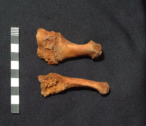
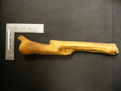
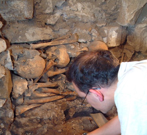
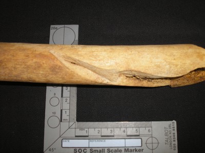
Archaeology is not just digging up bones and other artefacts. Sometimes, it can throw up some interesting ‘whodunit’ questions. This post reports on just such an instance. The osteaoarchaeologist, Paul Duffy, while carrying out the detailed analysis of the bones uncovered during the archaeological dig in 2006, came across severe injuries on the upper left leg of a man. From the evidence he was aged perhaps in his early 30s. A view of the lower part of his femur, just above the knee, is shown in the first photograph.

A general view of the badly injured femur

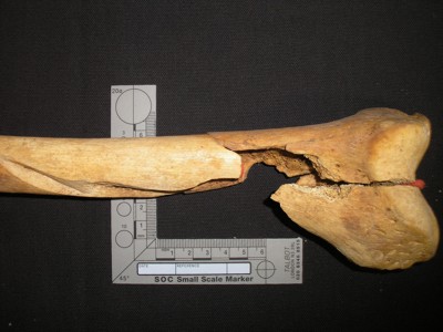
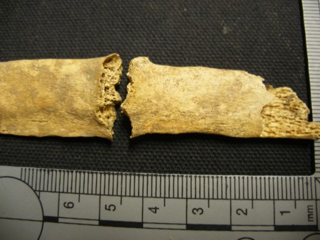
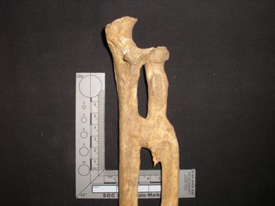
The most obvious thing is major damage and breaking of the bone. Additionally there had been no healing of the fractures, which suggests that he died around the time the injury was inflicted. A closer look shows that there is a deep blade wound (shown in close-up in the second photograph).

A closeup of the ‘chop’ wound


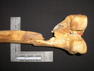
This damage is characteristic of a ‘chop’ type of injury. This would also have severed blood vessels, muscles, tendons and nerves and no doubt there would have been severe bleeding. This man would have experienced extreme shock. Together these factors could well have been the cause of his death.

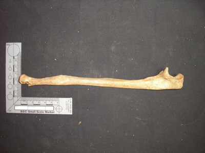
So how did this happen? The third photograph shows a different view of the same injury where the shattering of the lower part of the femur is more obvious.

The same bone from the opposite side

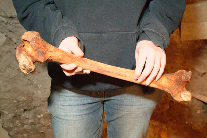
This would have required considerable force and would have come from behind. A ‘reconstruction’ is shown in the last photograph indicating that the injuries are consistent with a blow to the lower part of the upper leg. There is, obviously, no direct evidence, but it is most likely that the unfortunate man was attacked from the rear, perhaps as he was running away from a would-be attacker or a fight or battle.

A reconstruction of how the injury might have been inflicted


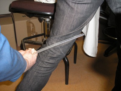
We are indebted to Paul Duffy for the detective work – and all the studies on the bones. No researcher was injured in the reconstruction! Paul is director of Brandanii Archaeology and Heritage, Bute, which can be accessed on Twitter and Facebook at present whilst their website is refreshed. The photographs are copyright Aberdeen Art Gallery & Museums Collections and are used with permission.

Prostate Cancer

There is a tendency to think that cancer is a relatively new disease, but this is not the case. It is certainly true that there has been a growth in its incidence in the ‘developed world’, but whether this is due to better diagnosis or to lifestyle is a moot point. It may not have been expected that the archaeological record would produce evidence of cancer. However, the human remains i.e. bones, found during the archaeological dig in the former East Kirk during 2006 do show a few cases of cancer affecting bone.

The prostate gland is a small gland located just below the outlet of the bladder in males. The prostate gland was first described anatomically in 1536. Prostate cancer was not specifically recorded until 1853. Prostate cancer occurs when a growth starts within the gland. In most cases the only problem it is likely to cause is with urinating. However, like other cancers, it can spread outside the prostate into the lymph or metastasise into bone with the back and the hip the common sites of these bony metastatic tumours. It is at this stage that this cancer can become a threat to life.

post-78-a-case-of-prostate-cancer-on-pelvic-bone-spicules-upper-left
Part of the pelvis of an old man showing evidence of prostate cancer

The photograph shows part of the pelvis of an older man. Towards the top left of the bone are ‘spicules’ – the roughened area – which are characteristic of metastatic prostate cancer. It is likely that he developed considerable pain in his pelvis as a result, a common indication of progression of prostate cancer. We do not know whether this is what caused his death. The date when this man died is not known, but it was somewhere between 15th and 18th century, so was well before the disease was recognised.


(The photograph is copyright Aberdeen Art Gallery & Museums Collections and is used with permission).

Perthes Disease

It is surprising how many diseases, even relatively uncommon ones, can be identified in human remains uncovered during an archaeological dig. That was particularly true of the large dig which took place in the former East Kirk of St Nicholas in 2006. This blog deals with one of these less common conditions – usually known as Perthes Disease, but more properly known as Legg-Calve-Perthes disease (after the three doctors who identified the condition). The medical term is avascular necrosis.

The condition arises in childhood, between the ages of about 2 and 10, with the highest incidence in the range of 4-8 years. At this age, the bones are still growing, so anything which interferes with this is likely to cause problems. Perthes disease affects the head of the femur – the ball of the thigh bone which fits into the hip. For a reason which is not understood, there is an interruption of the blood supply to this actively growing region of the bone. That causes the bone cells to die – that is where the term avascular necrosis comes from. So the bone stops growing and there is a loss of bone mass resulting in a weakness and wearing away of the bone. At this stage a child will suffer aching in the hip (similar to ‘growing pains’) and may develop a limp. However, with time the blood supply re-establishes and the bone starts growing again. But damage will probably have been done to the bone during the phase when it was dead, often a change of shape will have occurred, so a deformity is a common outcome.

E84 St Nicholas Site Week of 05.06.06 Gemma holding diseased bone Aberdeen Art Gallery & Museums Collections
Damage to the head (ball) of the femur – seen on the lower left

The photograph shows one of the examples found in the dig. This is of an adult man – it is more common in men – and the deformity of the ‘ball’ is quite clearly seen. The hip end of the femur is to the left, and the knee end to the right. At the head end of the bone, the ‘ball’ is pointing downwards. Obviously this is supposed to be round, but it clearly is not. The consequences for the sufferer is an initial shortening of the leg (it usually only affects one leg) giving a pronounced limp and there could be quite limited movement depending on the severity of the deformity. Longer term it is likely that osteoarthritis will develop with all the pain and difficulty which that will bring.

We are grateful to Paul Duffy, osteoarchaeologist, for the detailed studies on the human remains (http://www.discoverbutearchaeology.co.uk/).

(The photograph is copyright Aberdeen Art Gallery & Museums Collections and used with permission.)

Ankylosis

Ankylosis is the medical term used to describe an abnormal adhesion and rigidity of the bones in a joint. The potential causes are many, but frequently arise from injury or disease. Initially in ankylosis there is inflammation in the joints affecting the tendons, muscles and the membranes lining the surfaces of the bones. This is often associated with swelling and pain. The consequence of this is that the bones do not move easily and so they tend to become fixed in one particular position, which may be the one which causes least pain. However, this immobility can then lead to the bones fusing together. When this has happened no movement is possible in the joint, although surgical intervention can sometimes re-establish movement, such as with an elbow or shoulder. There are several different types of ankyloses. One of the common forms arises when the person has an inflammatory disease, such as rheumatoid arthritis which most often affects the fingers and wrist. During the archaeological dig in the former East Kirk a number of examples of ankylosis were found among the human remains, but they were affecting the feet.

E84 St Nicholas Site Week 36 Foot bones from SK665 Aberdeen Art Gallery & Museums Collections
Two metatarsal bones (to the right) fused to two tarsal bones (to the left)

The accompanying photographs show two examples of the completely fused bones in the foot which were found. The first one shows two metatarsals (the long bone in the mid-foo, to the rightt) fused to tarsal bones (in the ankle, to the left), whilst the second photograph shows a toe.

Post 71 (b) July 07 Toe - end two bones fused - was found commonly in dig
A toe showing the bones completely fused together


There is a tendency to imagine that this type of disease is ‘modern’ and confined to humans. The examples shown were of individuals who lived several hundred years ago. However, paleopathologists have found evidence of ankylosis in the fossil record, including one example of the famous dinosaur Tyrannosaurus rex!

(The photographs are copyright Aberdeen Art Gallery & Museums Collections and are used with permission).

Broken bones

Broken bones happen! They are a nuisance and can be painful, but given correct medical input and with time, they will heal. The detailed healing process is fairly complex and occurs in distinct stages. The first of these is the ‘reactive stage’ when there is an inflammatory response and the start of laying down new cells. This stage normally lasts for 3-5 days. Next is the ‘reparative stage’ when cartilage and then bone cells are laid down, a process which can take up to 12 weeks, but can be as short as 2 weeks. Finally there is the ‘remodelling stage’ during which the original bone structure is fully restored. The duration of this final stage is very variable and can take several years to complete. The time taken to heal varies with the location of the bone (a finger takes about 2 weeks, whilst the femur can be about 12 weeks) but it will also depend on many other factors such as age, smoking, nutrition, underlying diseases and related drug treatments. Medical intervention is normally fairly straightforward and involves ‘pushing’ the bones back in place and then stabilising them during the natural healing process.


During the archaeological dig in the former East Kirk of St Nicholas a large number of human remains were found – as expected in what had been a graveyard before the building was extended over it. Amongst these, several examples of fractures in different states of healing were found. We are grateful to Paul Duffy of Brandanii Archaeology for doing the detailed analysis and taking the photographs. A few examples are shown here to illustrate some of the different situations which were found.

Post 67 (a) Unhealed at time of death rib fracture in old man
No healing of fractured rib

In the first example, a rib has been broken and no healing has taken place. Obviously we do not know why it was broken, but the break must have occurred a fairly short time before death. It could even have been an injury received at the time of death.


OLYMPUS DIGITAL CAMERA
Well healed bone


The second example is of a bone which had been broken but which has healed. The ‘remodelling phase’ is not quite complete as the bone has not regained its normal profile – there is still some callous causing a small budge at the site of the fracture.


Post 67 (c)  Mis-aligned heal of tibia
Shortened leg due to bad ‘setting’

The third example is of a fracture which has fully healed but, as can be clearly seen, the bone was not ‘set’ properly before healing took place. The two halves have fused together and there is a smooth profile to the bone. This is a tibia (lower leg bone), which means that this person would have had one leg shorter than the other by three inches or more.


OLYMPUS DIGITAL CAMERA
Lower arm bones fused preventing rotation


The final example is of the lower forearm. The breaks here were not set properly, if at all. However, the bones have fused – but in two separate places. The normal rotational movement of the lower arm is possible because the two bones, the radius and ulna, can rotate around each other. This unfortunate person would have lost that ability and would have been left with very little useful movement in that arm.

Syphilis

A great deal about the health of individuals interred at St Nicholas Kirk can be learned from their remains uncovered during the archaeological dig. This is one example, others will also be featured, which perhaps comes as a surprise.

From both archaeological and written records, it appears that syphilis was unknown in Europe until the 1490s. It rapidly became a common venereal disease. It is caused by a micro-organism called Treponema pallidum – a spirochete for those interested. An infection can pass through three distinct stages if untreated. The initial stage is of a skin lesion at the site of infection – primary syphilis. Secondary syphilis occurs up to 6 months later and is characterised by a general rash. It may then enter a dormant phase which can last for several decades before entering the final tertiary phase. In this there are soft tissue swellings, called gumma, but these cannot be seen in archaeological specimens. However, there can be changes to the bone and this is how we know that there was at least one case of syphilis uncovered during the archaeological dig in the former East Kirk in 2006.

Post 52 Skull showing clear evidence of syphilis
Skull of a man who had tertiary syphilis

The photograph is of the skull of a person, probably male, aged between 17 and 25 years old. On it can be seen the characteristic lesions caused by tertiary syphilis. These lesions start as a small depression on the skull, called Caries Sicca, associated with inflammation in the tissue immediately in contact with the bone. This gradually deepens and widens and later new bone is deposited around the edge, giving a slight ridge (called a periosteal reaction). There can be similar reactions on other bones of the body. There were such lesions on all the ‘long bones’ of this person, but these lesions are not as easy to see or as distinctive. It can only be speculation, but the feeling is that whilst this young person could have acquired the infection through his own actions, it is equally possible that this is a case of so called congenital syphilis; in other words, his mother infected him before or during his birth.

We are very grateful to Dr Paul Duffy, our human bone specialist during and following the dig, for his detailed analysis. (His website is http://www.discoverbutearchaeology.co.uk/) The photograph is copyright Aberdeen Art Gallery & Museums Collections and is used with permission.

Bones in walls!

During the archaeological dig in 2006 there were a number of places where bones were found either under a wall or built into a wall. One reason for this arises because the walls were built in what had been, up to that time, the graveyard – in other words the graves already existed and the wall was simply built on top.

E84 St Nicholas Site Week 28 Skeleton in wall - SK426 Aberdeen Art Gallery & Museums Collections
Bones under the 15th century wall

The first two photographs show examples of this under the 15th century wall. The first photograph clearly shows how the wall was built on top of the burial. In the second photograph, there is also a bone higher up the wall.


Post 47 (b) bones DSCF0169 Bones in exterior (south) wall
Bones under and in 15th century wall

When the bones are built into the wall they would have been placed there deliberately when building was taking place. Any soil disturbance in the graveyard could disturb existing burials. So digging a new grave was likely to uncover some remains. Likewise, when foundations were being dug the same could happen. In the latter situation it was normal to incorporate the bones within the wall. This might seem strange to modern minds, but bone is a very strong material and so could be used in this way (the first two photographs show bones more than 500 years old and still supporting the wall).


E84 St Nicholas Site Week 35 Stewart (Robbo) with skeletons in wall (Ian's section) Aberdeen Art Gallery & Museums Collections
Large collection of bones built into a wall

The third photograph shows a much larger assemblage of bones found in a wall towards the west end of the dig area. Presumably several graves were disturbed during site preparation. The archaeologist is actually working on a grave right at the foot of the wall.


Post 47 (d) DSCN0446
Bones sticking out of a 19th century wall


The final photograph shows a wall, which still exists, which was probably built when the East Kirk was rebuilt in 1837. It shows some of the bones which stick out from the wall. There are many more, including one or two skulls in the whole wall.


First three photographs are copyright and used with permission of Aberdeen Art Gallery & Museums Collections