Broken bones happen! They are a nuisance and can be painful, but given correct medical input and with time, they will heal. The detailed healing process is fairly complex and occurs in distinct stages. The first of these is the ‘reactive stage’ when there is an inflammatory response and the start of laying down new cells. This stage normally lasts for 3-5 days. Next is the ‘reparative stage’ when cartilage and then bone cells are laid down, a process which can take up to 12 weeks, but can be as short as 2 weeks. Finally there is the ‘remodelling stage’ during which the original bone structure is fully restored. The duration of this final stage is very variable and can take several years to complete. The time taken to heal varies with the location of the bone (a finger takes about 2 weeks, whilst the femur can be about 12 weeks) but it will also depend on many other factors such as age, smoking, nutrition, underlying diseases and related drug treatments. Medical intervention is normally fairly straightforward and involves ‘pushing’ the bones back in place and then stabilising them during the natural healing process.
During the archaeological dig in the former East Kirk of St Nicholas a large number of human remains were found – as expected in what had been a graveyard before the building was extended over it. Amongst these, several examples of fractures in different states of healing were found. We are grateful to Paul Duffy of Brandanii Archaeology for doing the detailed analysis and taking the photographs. A few examples are shown here to illustrate some of the different situations which were found.
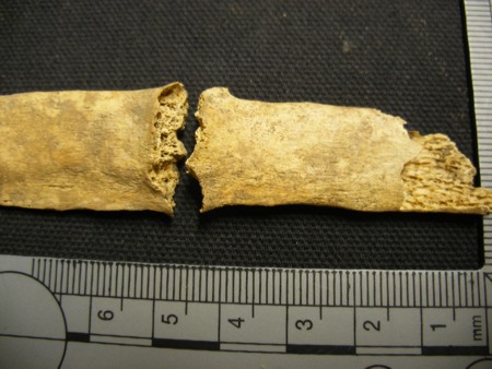
In the first example, a rib has been broken and no healing has taken place. Obviously we do not know why it was broken, but the break must have occurred a fairly short time before death. It could even have been an injury received at the time of death.
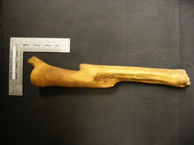
The second example is of a bone which had been broken but which has healed. The ‘remodelling phase’ is not quite complete as the bone has not regained its normal profile – there is still some callous causing a small budge at the site of the fracture.
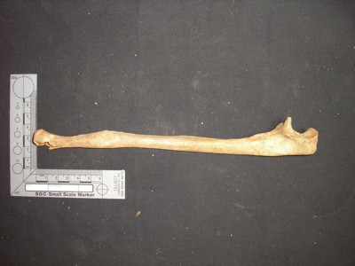
The third example is of a fracture which has fully healed but, as can be clearly seen, the bone was not ‘set’ properly before healing took place. The two halves have fused together and there is a smooth profile to the bone. This is a tibia (lower leg bone), which means that this person would have had one leg shorter than the other by three inches or more.
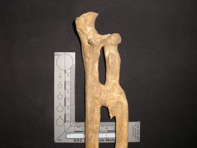
The final example is of the lower forearm. The breaks here were not set properly, if at all. However, the bones have fused – but in two separate places. The normal rotational movement of the lower arm is possible because the two bones, the radius and ulna, can rotate around each other. This unfortunate person would have lost that ability and would have been left with very little useful movement in that arm.